Image:Diatoms.png
Un article de Wikipédia, l'encyclopédie libre.
Taille de cet aperçu : 734 × 600 pixels
Image en plus haute résolution (1 400 × 1 144 pixels, taille du fichier : 951 Kio, type MIME : image/png)
| | Ce fichier provient de Wikimedia Commons?. Les informations le concernant sont affichées ci-dessous (procédure). |
| Description |
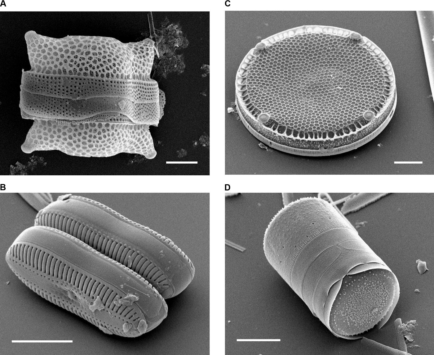
Scanning Electron Micrographs of Diatoms. (A) Biddulphia reticulata. The whole shell or frustule of a centric diatom showing valves and girdle bands (size bar = 10 micrometres). (B) Diploneis sp. This picture shows two whole pennate diatom frustules in which raphes or slits, valves, and girdle bands can be seen (size bar = 10 micrometres). (C) Eupodiscus radiatus. View of a single valve of a centric diatom (size bar = 20 micrometres) (D) Melosira varians. The frustule of a centric diatom, showing both valves and some girdle bands (size bar = 10 micrometres). |
||||
|---|---|---|---|---|---|
| Source |
Bradbury J: Nature's Nanotechnologists: Unveiling the Secrets of Diatoms. PLoS Biol 2/10/2004: e306. http://dx.doi.org/10.1371/journal.pbio.0020306 |
||||
| Date |
Published: October 12, 2004 |
||||
| Author |
Images courtesy of Mary Ann Tiffany, San Diego State University. |
||||
| Permission (Reusing this image) |
|
Historique du fichier
Cliquer sur une date et une heure pour voir le fichier tel qu’il était à ce moment-là
| Date et heure | Dimensions | Utilisateur | Commentaire | |
|---|---|---|---|---|
| actuel | 16 novembre 2006 à 20:10 | 1 400×1 144 (951 Kio) | Ayacop | ({{Information |Description='''Scanning Electron Micrographs of Diatoms.''' (A) ''Biddulphia reticulata''. The whole shell or frustule of a centric diatom showing valves and girdle bands (size bar = 10 micrometres). (B) ''Diploneis sp.'' This picture shows) |
Pages contenant l’image
Les pages ci-dessous contiennent cette image :

